|
Home |
|
Research |
|
Publications |
|
Lab members |
|
Collaborators |
|
Teaching |
|
Research opportunities |
|
Contact us |
|
Fun Pics |
|
Wylie Lab |
|
Research |
|
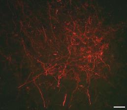
Neuroanatomy and Neurochemistry of Brainstem-Cerebellar Pathways We have been using a variety of techniques to investigate the neuroantomy and neurochemistry of brainstem cerebellar pathways that are involved in the processing of optic flow. To investigate the intricacies of the connections, we use biotinylated dextran amine (BDA) and cholera toxin subunit B (CTB) as anterograde and retrograde tracers, as well as fluorescent tracers. Also, we investigate resident neurochemical composition with fluorescent microscopy .
Fluorescently labeled BDA terminals in the inferior olive from injections in nBOR
Specific lines of research:
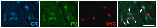
Projections of the nucleus of the nucleus of the basal optic root (nBOR) and pretectal nucleus lentiformis mesencephali (LM) in Pigeons: The nBOR and LM are the retinal-recipient nuclei in birds that process optic flow information. Using anterograde and retrograde tracers we have been examining the projections of LM and nBOR to various target structures (Wylie et al., 1997; 1998; 1999; 2005; Wylie and Linkenhoker, 1996; Wylie, 2001; Pakan et al., 2006a,b; Winship et al., 2006). We have also been pursuing work in attempt to determine the neurotransmitter profiles of different cell types within the LM and nBOR (Winship et al., 2006; Pakan et al., 2006a).
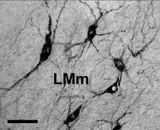
VbC projecting neurons in LM
Calcium binding proteins (Calretinin: CR; Parvalbumin: PV) and VbC projecting cells in nBOR
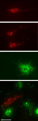
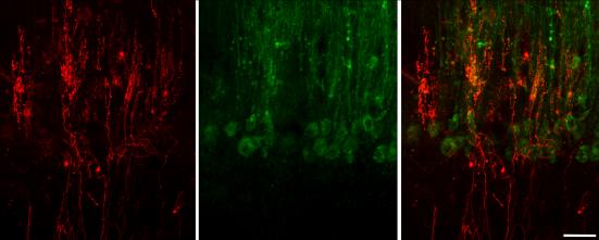
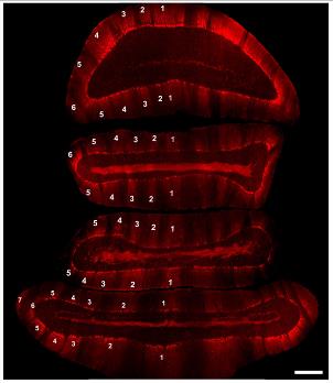
Functional Neuroanatomy of the Inferior Olive (IO) - Vestibulocerebellar (VbC) System: The LM and nBOR project to parts of the inferior olive (IO), which in turn project to to the VbC as climbing fibres. VbC Purkinje cells respond to particular patterns of optic flow that result from either self-translation or sefl-rotation. The different types of neurons are topographically organized into parasagittal zones, typical of cerebellar organization. We have been examining the connectivity of these zones using anterograde and retrograde tracers. In collaboration with Dr. Richard Hawkes at the University of Calgary, we have also been examining how the different optic flow zones in the VbC organization relates to zones revealed with the molecular marker Zebrin II. Zebrin II is expressed in by some Purkinje cell, revealing a sagittal striping pattern. These may correlated with the topography of optic flow neurons in the VbC.
Climbing fibres (red) and zebrin II labeling (green) in the VbC
Pattern of zebrin zones in the pigeon cerebellum, creating a “striped” appearance. |
|
Dept. of Psychology & Neuroscience and Mental Health Institute (MMHI) |