
|
Home |
|
Research |
|
Publications |
|
Lab members |
|
Collaborators |
|
Teaching |
|
Research opportunities |
|
Contact us |
|
Fun Pics |
|
Wylie Lab |
|
Research |
|
Neurophysiology of Motion Processing in the Avian Brain A major focus of my research concerns the neurophysiological basis of visual processing, in particular those parts of the brain involved in the processing of optic flow that results from self-motion. Because the world consists of stationary objects and surfaces, self-motion through the environment induces patterns of motion across the entire retina, known as optic flow.
Optic flow provides a rich source of proprioceptive information, and can be used for several behaviors including determination of heading, control of posture and locomotion, perception of self-motion and navigation.
Optic flow occurs across the retina as we move through our environment.
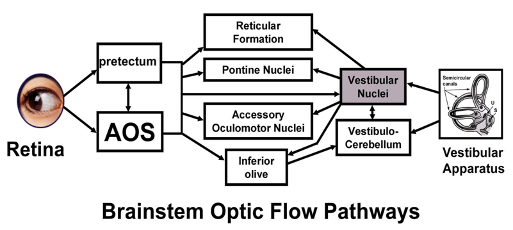
Optic flow is analyzed by two specific visual pathways, a pretectal pathway and the Accessory Optic System (AOS), and is integrated with vestibular information in the vestibulocerebellum (VbC). We know that the these pathways to the cerebellum are critical for controlling the optomotor or optokinetic response. The optokinetic response is ubiquitous to the animal kingdom and can be demonstrated in both vertebrates and invertebrates. Gaze stabilization is critical for normal visual function: without gaze stabilization visual acuity is dramatically impaired as is velocity discrimination. Damage to the AOS and pretectum, but not other visual pathways, severely compromises or even abolishes the optokinetic response.
Basic pathways that analyze optic flow and control the optokinetic response
Specific lines of research include:
Self-Translation vs. Self Rotation: Most of my work has shown that the nucleus of the basal optic root (nBOR) in pigeon AOS, the pretectal nucleus lentiformis mesencephali (LM) and the VbC, contain neurons that are specialized for processing particular patterns of optic flow that result from either self-rotation or self-translation. In this body of work we stimulate neurons with optic flow patterns produced by planetarium projectors. Some of my most significant findings in this regard, published in Nature, illustrated that the self-translation and self-rotation systems share a common frame of reference with the vestibular canals and eye muscles (Wylie, Bischof and Frost, 1998).
Early motion processing in the Accessory Optic System: In another line of research, we have been investigating early motion processing in the nBOR and LM. For example, using drifting sine wave gratings as stimuli, we have found that there are two groups of neurons in both the LM and nBOR (Crowder and Wylie, 2001; Wylie and Crowder, 2001). "Fast neurons" respond best to stimuli of low spatial and high temporal frequency, whereas "slow neurons" prefer high spatial and low temporal frequency. It remains to be seen how these different groups contribute to the optokinetic reflex.
Comparative Perception: In collaboration with Dr. Marcia Spetch, who is an icon in the field of comparative cognition, I have been investigating the perceptual abilities of pigeons and attempting to determine their neural correlates. In these studies we are evaluating evolutionary hypotheses and the neural explanations of perceptual behaviors in humans and other species. For example, in pigeons, we have determined the thresholds of motion perception in random dot displays varying in coherence , and the thresholds for detection of spatial patters (for example, detection of gratings in static noise). The Ectostiatum in pigeons is thought to be homologous to the cortical area MT in mammals. In monkeys, lesions to MT increase the thresholds for motion perception in these displays. Lesions to the caudal part of ectostriatum have a similar effect in pigeons, whereas lesions to the rostral part of Ectostriatum disrupt spatial vision (Nguyen et al., 2004). |
|
Dept. of Psychology & Neuroscience and Mental Health Institute (MMHI) |
|
© Doug Wylie, all rights reserved All images are copyrighted and may not be used in any way. |