|
Home |
|
Research |
|
Publications |
|
Lab members |
|
Collaborators |
|
Teaching |
|
Research opportunities |
|
Contact us |
|
Fun Pics |
|
Wylie Lab |
|
Research |
|
Comparative Neuroanatomy: In collaboration with Dr. Andrew Iwaniuk, Lethbridge University. We have been fortunate to obtain a wealth of brains from over 100 different species of birds from numerous museums and private collectors. With these data were are analysing the size of various nuclei in the brain and attempting to correlate this with behavioural differences amongst various bird species. We have been looking at the size of various nuclei in the avian brain, including the visual areas in the brainstem and cerebellum. Thus, this comparative work nicely complements my neurophysiological and neuroanatomical work.
Specific lines of research:
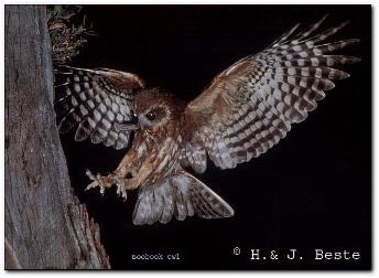
Size of the MLd across species: The nucleus mesencephalicus lateralis dorsalis or MLd is homologous to the inferior colliculus of mammals and it critical for sound localization. We found that the MLd is significantly enlarged in owls (Iwaniuk et al., 2006). This likely reflects the uncanny ability of some owls to locate prey using only auditory cues. Interestingly, the enlargement of MLd was not uniform in the owls that we examined. Asymmetrically eared owls, such as the Northern Saw-whet Owl (Aegolius acadicus) and Barn Owl (Tyto alba), have much larger MLd's than symmetrically eared owls, such as the Southern Boobook Owl (Ninox boobook). How this enlargement might be related to the degree of ear asymmetry within owls is currently under investigation.
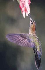
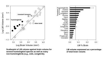
The Pretectal Nucleus Lentiformis Mesencephali is Hypertrophied in Hummingbirds: I have been studying in the neuroanatomy and neurophysiology of the nucleus lentiformis mesencephali, or LM, for quite some time. We know that the LM is involved in the analysis of optic flow and the generation of the optomotor response to facilitate gaze stabilization . Recently, Andy and I examined variation within the LM in birds. Of all of the feeding methods known to occur in birds, hovering flight is perhaps the most demanding in terms of gaze stabilization. Hummingbirds and other hovering species (e.g., sunbirds, kingfishers, raptors) need to keep their vision stable in order to successfully feed. After examining the brains of several hummingbird species, we found that LM was significantly enlarged in hummingbirds compared to other birds. Other species that hover, such as the Eastern Spinebill (Acanthorhynchus tenuirostris), American Kestrel (Falco sparverius) and Belted Kingfisher (Ceryle alcyon) also had an enlarged LM, but not to the same extent as the hummingbirds. In contrast,other visual structure including the nucleus of the nasal optic root (nBOR), the ventral leaflet of the lateral geniculate nucleus (GLv) and the optic tectum (TeO) were all not significantly larger in hummingbirds. Thus, the hypertrophy is specific to LM and is present in all hovering species, but greatest in the hummingbirds.
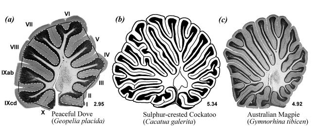
Variation in Cerebellar Morphology Among Birds: More recently, we have begun to use sophisticated multivariate methods as well as discriminant function analysis to explore the evolution of the cerebellum in birds (Iwaniuk et al., 2006a, 2006c,). In these analyses, we have shown that the degree of folding of the entire cerebellar cortex is dependent primarily upon body size, brain size and phylogenetic history. In fact, significant differences in the degree of cerebellar foliation are clearly evident among orders. Parrots and seabirds, for example, have highly folded cerebellar whereas pigeons and chicken-like birds do not. The proportional size of individual folia is also dependent on body size, brain size, phylogenetic history, but also, to some extent, behaviour. Species classified as strong fliers, such as hummingbirds, swifts and raptors, all share significanly larger folia VI and VII compared to other birds. Although this might reflect wing projections to and from the cerebellum, we suspect that this may actually reflect visual input to the cerebellum that is critical for steering behaviour. Currently, we are addressing whether these changes in the size of the folia also reflect differences in the size of cells in the Purkinje, granule and molecular cell layers of the cerebellum as well as correlated changes among folia and extra-cerebellar regions of the brain.
|
|
Dept. of Psychology & Neuroscience and Mental Health Institute (MMHI) |