Main Page
![]()

![]() Lecture Notes
Lecture Notes
![]()
![]() Chapter 4
Chapter 4
Chapter 4 (and Related) Lecture Notes
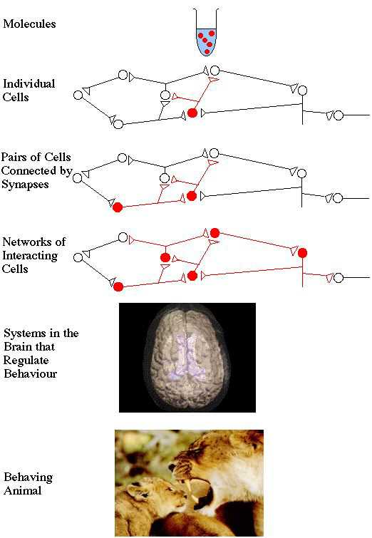
Levels of Neuronal Organization
Neurons
- Signaling
- Information transfer
- Within cells
- Between cells
- Neuroglia
- Astrocytes
- Myelin
- Oligodendrocytes
- Schwann cells
- Neurons
- Sensory
- Interneuron
- Motor
- Soma
- Axon
- Terminals
- Dendrites
Soma
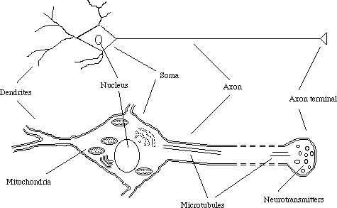
Like any other cell
- Nucleus, mitocondria, plasma membrane
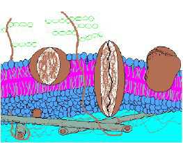
plasma membrane
Axon
- Communication
- Connect to:
- Dendrites
- Soma
- Oligodendrocytes (CNS)
- Schwann cells (PNS)
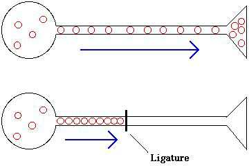
- Microtubules ("axon escalators")
- Cytoskeleton
- Neurotransmitters
- Proteins
Axon Terminal
Connects with:
- Dendrites (common)
- Soma (common)
- Other axons (rare)
- Chemical transmitters
- Neurotransmitters
- Electrical signals
- Excitatory or inhibitory signals
Dendrites
Connects to axon terminals
Transfers signals received from another neuron to the soma
Axons and dendrites form synapses!
The Synapse
- Presynaptic cell
- Transmitter
- Postsynaptic cell
- Receiver
- Chemical and electrical synapses
- Demonstrates chemical (neurotransmitter) synapse
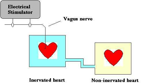
Chemical Synapses
- Presynaptic
- Axon terminal (contains neurotransmitter)
- Postsynaptic
- Dendrite or soma
- Space between pre- and postsynaptic elements
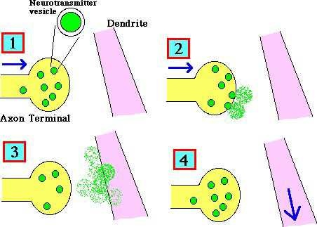
- Axon terminal releases neurotransmitter
- Neurotransmitters diffuse across synaptic cleft
- Postsynaptic receptors uptake neurotransmitter
- Signal travels to next neuron (unidirectional!)
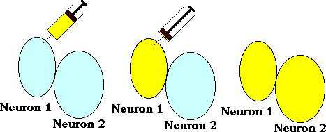
Electrical Synapses
Pre- and postsynaptic elements physically indistinguishable
Bi-directional!
Gap junctions
- Cell-to-cell pores
- Mediates signalling
- Low resistance pathways
- Neurons are physically coupled
- No synaptic cleft to diffuse across
- Lucifer yellow
Membrane Potential
Resting potential
- -40mV to -90mV
- Negative relative to extracellular
Depolarized
- Less negative than resting potential
- More negative than resting potential
Action Potential
A large depolarization of the axon
- "Firing" of the axon
- Critical point
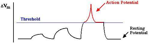
All or none
- Either threshold is reached, or nothing happens
- Can't stop an action potential
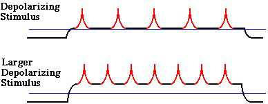
Frequency coding
- Larger depolarizing stimulus, greater firing rate
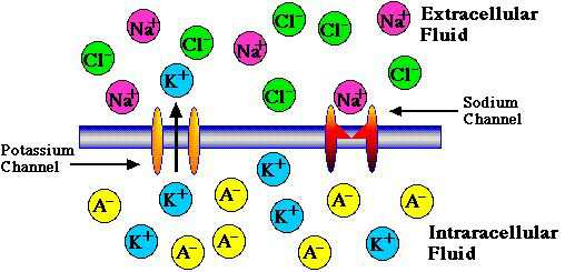
Molecular Basis for Action Potentials
- Porous skin of neuron
- Intracellular fluid
- Extracellular fluid
- Ion channels
- Potassium
- Sodium
- Balance of charge
- Intracellular more negative than extracellular
- Resting potential (negative)
Action Potential
- Initiation phase
- Sodium channels open
- Depolarization phase
- Sodium ions enter
- Concentration gradient
- Electrical gradient
- Repolarization phase
- Sodium channels close
- Potassium channels open
- Potassium ions exit
- Sodium-potassium pump
- Sodium ions enter
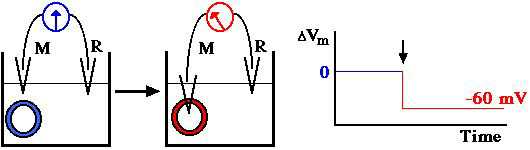
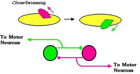
Central Pattern Generators
- e.g., walking, swimming, peristaltic movement
- Contract, relax, contract, relax...
- Different neurons innervate different muscles
- Two neurons
- Reciprocal inhibition
- Postinhibitory rebound
- Cell becomes more excitable after hyperpolarization (threshold is reduced)
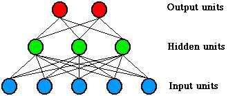
Artificial Neural Networks (ANNs)
Must be trained
- Learning based on neuronal structure
- Input units
- Sensory neurons
- Hidden units
- Interneurons
- Output units
- Motor neurons
- No
- Simulation
- Metaphore
- Hypothesis testing
Nervous System Organization
- Brain
- Spinal cord
- Non-regenerative
- Nerves outside the CNS
- Regenerative
- Sensory neurons
- Carry information from sensory organs to CNS
- Interneurons
- Only in CNS
- Carry information from one neuron to another
- Organize and integrate information
- Motor neurons
- Carry information to muscles and glands
Peripheral Nervous System
- Muscles attached to bones
- Externally observable movements
- Neurons extend directly from CNS to muscle
- Visceral muscles and glands
- Indirect neuronal action from CNS to ganglion to target site
- Sympathetic division
- Immediate responses to stressful stimuli
- Parasympathetic division
- Regenerative, energy-conserving processes
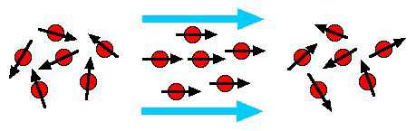
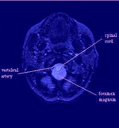
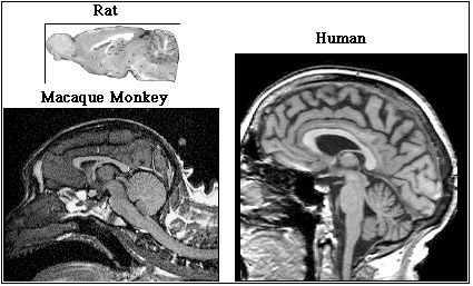
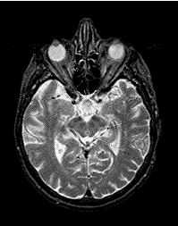
Magnetic Resonance Imaging (MRI)
- Hydrogen dipoles
- Normally randomly positioned
- Repositions hydrogen dipoles
- Takes time for dipoles to re-position after magnetic field turned off
- Long axis
- 100 to 2000 ms in biological tissue
- Perpendicular to long axis
- 30 to 300 ms in biological tissue
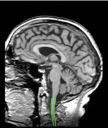
Central Nervous System
- Ascending tracts
- Somatosensory information to brain
- Descending tracts
- Motor-control information from brain
- Central pattern generators
- Interneurons
- Brain influences
- Spinal reflexes
- Sensory neuron
- Interneuron
- Motor neuron
- e.g., flexion reflex
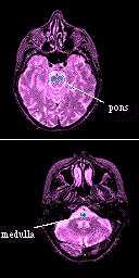
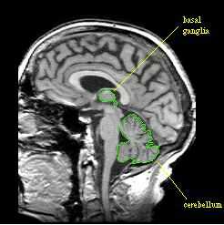
Subcortical Structures of the Brain
- Cranial nerves
- Postural reflexes
- Balance
- Vital reflexes
- Breathing, heart rate, etc.
- Mid-brain
- Basic movements
- Connections to CPGs in spinal cord
- Thalamus
- "Relay station"
- Connects parts of the brain
- Sensory tracts from brainstem terminate here
- Output then to cerebral cortex
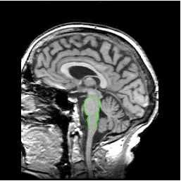
Cerebellum
- Motor control
- Rapid, powerful movements
- Information processor
- Input from sensory organs
- Coordinates complex movement
- Grey matter
- Unmyelinated
- Motor control
- Slow, precise movements
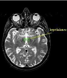
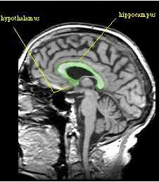
Limbic System and Hypothalamus
- Amygdala
- Scent
- Drives and emotion
- Hippocampus
- Memory hypothalamus
- Regulates internal environment of the body
- Autonomic nervous system
- Hormones
- Drive states
Cerebral Cortex
- Input from sensory nerves (via thalamus)
- Visual area
- Auditory area
- Somatosensory area
- Back
- Sends axons to motor neurons in brainstem and spinal cord
- Middle
- Perception, thought, decision making
- Receives input
- Sensory areas
- "Lower brain" structures
- Front
Cortical Asymmetry
Corpus callosum
- Connects hemispheres
- Sensory neurons from right side input to left hemisphere and vise versa
- Motor neuron from left hemisphere to right side of body
- Unified sensation and movement due to corpus callosum
- Left hemisphere
- e.g., Speech
- Right hemisphere
- e.g., Spatial tasks
Split Brain Patients
Contralateral pathways
Visual information
- Right visual field to left hemisphere
- Left visual field to right hemisphere
- Corpus callosum would transfer information
- Visual picture in left visual field
- Test: touch object with left hand
- Success!
- Test: name object
- Failure!
Hormones
- Related to neurotransmitters
Released by:
- Endocrine glands
- Internal organs
- Brain
Hormone effects
- Long-term
- e.g., Anatomical sex differences, growth, bone strength
- Short-term
- e.g., Fight-or-flight response, healing, menstrual cycle
- Peptides
- Pituitary gland
- Donąt pass cell membranes easily
- Act like neurotransmitters
- Open/close membrane channels
- Change ion concentrations
- Steroids
- Adrenal cortex and gonads
- Pass cell membranes easily
- Effect in cell nucleus
- Genes
- Production/reduction of certain proteins
- Pituitary
- Posterior
- Part of the brain
- Connected to neurosecretory cells in hypothalamus
- Produces hormone releasing factors
- Anterior
- Triggered by releasing factors
- Produces pituitary hormones
- Releases hormones into bloodstream
- Posterior